A spinal tumour can begin in the vertebrae, spinal cord coverings, or cord itself, or arrive there from a cancer in another organ. By the end, you will understand which symptoms need urgent assessment, how doctors confirm the diagnosis, and why treatment may involve surgery, radiotherapy, systemic cancer treatment, or a combination.
Key takeaways
- Seek assessment for persistent back pain, weakness, numbness, or bladder changes.
- MRI usually defines the tumour’s location; biopsy identifies its type when needed.
- Treatment may combine surgery, radiotherapy, chemotherapy, or targeted cancer treatment.
- Bring prior scans, biopsy reports, medication lists, and cancer records to your appointment.
What Is a Spinal Tumour, and Which Symptoms Need Assessment?
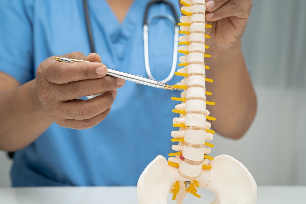
A useful spinal tumour definition is an abnormal growth arising in or near the spine, spinal cord, nerve roots, or supporting tissues. A primary spinal tumour begins in these structures; a metastatic spinal tumour spreads from another cancer, commonly in the breast, lung, kidney, or prostate.
In adults, a destructive lesion needs assessment for metastatic cancer, myeloma, or lymphoma rather than being assumed to be a primary tumour.
Location changes the likely diagnosis, surgical risk, and treatment options:
| Location | What it means | Common pattern |
|---|---|---|
| Vertebral tumour | Begins in a vertebral bone | Bone destruction, collapse, or instability |
| Epidural tumour | Lies outside the dura, often behind or beside the cord | Metastatic compression of the spinal cord or nerve roots |
| Intradural-extramedullary tumour | Sits inside the dura but outside the spinal cord | A mass displacing the cord or roots |
| Intramedullary tumour | Grows within the spinal cord | Cord expansion with progressive neurological symptoms |
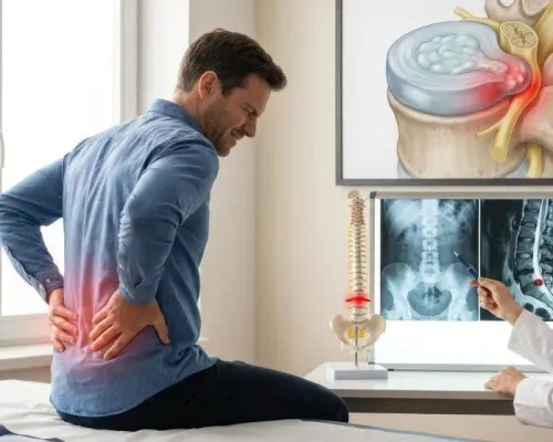
Arrange prompt medical evaluation for persistent back pain, pain that worsens at night, or pain wrapping around the chest or abdomen. Do not wait if you develop:
- Arm or leg weakness
- Numbness or loss of sensation
- Unsteady walking, frequent falls, or new gait change
- Loss of bladder or bowel control
Weakness, walking difficulty, or bladder and bowel changes require urgent assessment because spinal cord or nerve compression can cause permanent damage.
Evaluation usually combines a neurological examination, contrast MRI, CT for bone destruction, and staging scans such as PET-CT; biopsy is planned when imaging cannot establish the diagnosis or treatment would differ between tumour types, infection, and metastasis.
How Are Spinal Tumours Diagnosed and Biopsied?
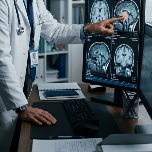
An MRI with and without contrast is the key test for locating a spinal tumour. It shows the spinal cord, nerve roots, epidural space, bone marrow and soft-tissue extension, while contrast helps distinguish active tumour from surrounding tissue. The scan also shows whether the cord is compressed and whether surgery may require decompression.
Tumour location narrows the likely diagnosis and affects the safest surgical route:
- Extradural: outside the dura, often involving vertebrae or spread from another cancer.
- Intradural-extramedullary: inside the dura but outside the spinal cord.
- Intramedullary: within the spinal cord itself.
A CT scan for bone destruction adds detail that MRI cannot match. It shows cortical erosion, calcification, fractures and the bony anatomy needed to plan fixation or reconstruction. PET-CT staging looks for a primary cancer elsewhere and for additional spread, especially when metastasis is suspected.
An image-guided biopsy provides tissue when scans cannot establish the diagnosis, no primary cancer is known, or treatment would differ between lymphoma, infection, metastasis and a primary spinal tumour. A pathologist uses histology, immunohistochemistry and, when indicated, molecular tests to identify the tumour’s origin, type and grade. The result guides surgery, radiotherapy and systemic treatment.
Biopsy is not required before every operation. Plan the needle route with the surgeon and radiologist so the tract can be removed during definitive surgery when appropriate.
How Do Doctors Choose Between Surgery, Radiotherapy, and Cancer Treatment?
The treatment plan turns on four findings: how much the spinal cord or nerve roots are compressed, whether the spine is mechanically stable, how the tumour responds to treatment, and how much cancer exists elsewhere.
| Factor | Findings that influence the choice | Likely treatment direction |
|---|---|---|
| Neurological compression | Progressive weakness, walking difficulty, bowel or bladder dysfunction, or high-grade metastatic spinal cord compression | Urgent decompression, often followed by radiotherapy |
| Spinal stability | Vertebral collapse, abnormal alignment, or severe movement-related pain | Stabilisation or reconstruction; a stable lesion may avoid open surgery |
| Tumour biology | Radiosensitive cancer, resistant tumour, or an uncertain diagnosis | Conventional radiotherapy, stereotactic body radiotherapy, biopsy, surgery, or a combination |
| Overall cancer burden | Widespread disease, limited life expectancy, or major medical frailty | Systemic cancer treatment, focused radiation, pain control, or supportive care rather than extensive surgery |
Radiotherapy suits many radiosensitive tumours and can control a painful, mechanically stable lesion. Stereotactic body radiotherapy delivers focused, high-dose treatment when precise targeting is safe; it does not replace urgent decompression or stabilisation.
Surgery becomes more compelling when neurological function is deteriorating, the spine cannot bear normal movement, tissue diagnosis is essential, or the tumour can be removed safely. Intramedullary tumours require particular caution because the surgeon must preserve functioning spinal-cord tissue. In widespread cancer, durable function and stability often matter more than complete tumour removal.
What Does Spine Tumour Surgery Aim to Do?
Surgery may seek a diagnosis, relieve pressure, remove tumour, or restore a spine weakened by disease. The right target depends on tumour location, biology, spread, neurological compression and spinal stability—not on removal at any cost.
| Option | What it means | When it applies |
|---|---|---|
| Biopsy-only surgery | Removes a tissue sample, not the tumour | Diagnosis is uncertain or treatment differs between metastasis, lymphoma, infection and a primary tumour |
| Spinal cord decompression | Removes tumour, bone or other tissue pressing on the cord or nerve roots | Weakness, walking difficulty, severe compression or threatened neurological function |
| Debulking surgery | Removes as much tumour as safely possible while leaving disease that is attached to critical structures | Full removal would cause unacceptable neurological injury |
| Subtotal resection | Deliberately leaves a measurable amount of tumour | Complete removal is unsafe or unnecessary because radiotherapy or systemic treatment can control the remainder |
| Complete resection | Removes all visible tumour within the planned surgical boundary | The tumour is localised and its position permits safe removal |
| Vertebral reconstruction | Rebuilds a destroyed vertebral body or spinal column using graft, cage or cement | Tumour has removed load-bearing bone |
| Fixation | Uses screws, rods or plates to hold unstable spinal segments | Disease or surgery has weakened alignment and mechanical stability |
Weigh the benefit against bleeding, infection, spinal cord or nerve injury, paralysis, cerebrospinal-fluid leak, hardware failure, persistent pain and tumour recurrence. A wider tumour resection can increase these risks, while inadequate decompression can leave neurological damage. In metastatic disease, preserving function and stability may matter more than radical removal.
Minimally invasive biopsy, decompression or percutaneous fixation can reduce muscle injury and recovery time, but it cannot eliminate tumour-related risks.
How Can You Prepare for Spine Tumour Care in Jaipur?
Before consenting to treatment, ask who will coordinate the case and what decision each specialist is making. A multidisciplinary spine tumour team should include a neurosurgeon in Jaipur, neuroradiology, pathology, radiation oncology, medical oncology and rehabilitation.
1. Who will review my MRI, CT and staging scans together? Ask whether the lesion is primary or metastatic, particularly if it is destructive and no breast, lung, kidney, prostate, thyroid cancer, myeloma or lymphoma has been identified.
2. Am I suitable for surgery now? Ask how spinal-cord compression, mechanical instability, tumour radiosensitivity, neurological function and your overall cancer burden affect the choice between biopsy, decompression, fixation, tumour removal, radiotherapy or systemic treatment. If Dr. Deepak Bariya is assessing you, ask which surgical aim is realistic: tissue diagnosis, nerve relief, stabilisation or resection.
3. Do I need a biopsy before surgery? It is important when scans cannot establish the diagnosis or treatment differs between lymphoma, infection, metastasis and a primary tumour. Ask the surgeon and radiologist to plan the biopsy route so the tract can be removed during definitive surgery when appropriate.
4. Which specialist owns the next treatment step? Confirm how pathology results will reach radiation oncology and medical oncology, and who will discuss radiotherapy, medicines or clinical surveillance.
5. What rehabilitation begins after treatment? Ask about walking, brace use, pain control, wound care, bladder or bowel symptoms and warning signs, then schedule the next review date and decision.
Related services
Brain tumours are one of the most critical neurological conditions, requiring accurate diagnosis, advanced surgical expertise, and timely treatment. View service → |
Headache And Back Ache Minimally Invasive Brain & Spine Surgery ₹500 What Is Minimally Invasive Brain & Spine Surgery? Minimally Invasive Brain & Spine Surgery (MIS) involves performing surgical procedures through very... View service → |
Frequently asked questions
What is a spinal tumour?
A spinal tumour is an abnormal growth in or near the spine, spinal cord, nerve roots, or supporting tissues. It may begin there or spread from another cancer.
Which symptoms need assessment for a spinal tumour?
Persistent or worsening back pain, pain that travels along a limb, weakness, numbness, walking difficulty, or new bladder and bowel problems need medical assessment.
How are spinal tumours diagnosed?
Doctors use a neurological examination and imaging, usually MRI. CT scans and other tests help assess bone involvement and the possible source of metastatic disease.
Why is a biopsy performed?
A biopsy examines tumour cells to identify the tumour type and guide treatment when imaging cannot provide a definite diagnosis.
How do doctors choose between surgery and other treatments?
The decision depends on tumour location, type, size, spinal stability, nerve or spinal cord compression, symptoms, overall health, and whether cancer has spread.
Related services
Endoscopic Spine Surgery( Slip Disc Surgery) What Is Endoscopic Spine Surgery? Endoscopic spine surgery is a minimally invasive procedure performed using a small endoscope—a thin tube with a... View service → |
Head And Spine Injury Treatment Head and spine injuries are medical emergencies that require immediate attention and expert care. View service → |
What Is Paralysis? Paralysis is the loss of muscle function in part of the body. View service → |
Brain Tumour And Hydrocephalus ₹500 What Is Endoscopic Brain Surgery? Endoscopic brain surgery is a minimally invasive procedure performed using an endoscope—a thin, flexible tube... View service → |