A brain haemorrhage is not one diagnosis, and the right treatment can range from intensive monitoring and medicine reversal to ventricular drainage, clot evacuation, aneurysm coiling or open surgery. You will learn how to respond before hospital, what scans reveal, which procedures fit different types of bleeding, and how to compare emergency capabilities in Jaipur.
Key takeaways
- Call emergency services for sudden weakness, speech trouble, collapse or severe headache.
- Keep the person still, protect their airway and give nothing to eat or drink.
- A CT scan identifies bleeding location, size and pressure on the brain.
- Treatment ranges from monitoring and medicines to clot evacuation or pressure-relief surgery.
Recognise the emergency and protect the patient before hospital
Call emergency services immediately if a person develops brain haemorrhage symptoms. A sudden severe headache, one-sided weakness, facial drooping and slurred speech can signal a stroke, and loss of balance, seizure, collapse, repeated vomiting or reduced consciousness also require urgent help.
1. Note the exact time symptoms began, or the last time the person was known to be well. Give this time to the emergency team.
2. Keep the person still and reassure them. If they are unconscious or vomiting but breathing, place them on their side and watch their breathing.
3. Do not give food, water, aspirin or any other medicine. Swallowing can fail after a stroke, and aspirin can worsen bleeding.
4. Gather the person’s current medicines, medical conditions and allergy information. Tell the emergency operator if they take warfarin, apixaban, rivaroxaban, clopidogrel or another blood thinner.
Do not drive the person yourself if an ambulance is available; emergency crews can begin assessment during transport. If the person stops breathing normally, follow the operator’s instructions for resuscitation. Do not assume improvement means the danger has passed: bleeding can expand quickly, raise pressure inside the skull and cause permanent injury.
Identify the type of bleeding and confirm it with urgent imaging
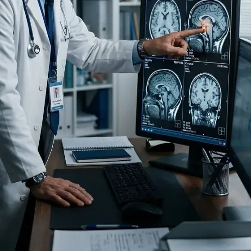
A non-contrast CT scan is usually the first decisive test. A haemorrhagic stroke shows fresh blood inside or around the brain; an ischaemic stroke results from a blocked blood vessel and may show little or no change during the first hours.
Symptoms alone cannot reliably distinguish them, so doctors arrange urgent imaging before giving treatments such as aspirin or clot-busting medicine.
The CT location and pattern help classify the bleed:
- Intracerebral haemorrhage: blood within brain tissue, often linked to high blood pressure, blood-thinning medicines, a tumour or a vascular abnormality.
- Subarachnoid haemorrhage: blood in the fluid-filled space around the brain, frequently caused by a ruptured aneurysm.
- Subdural haematoma: blood between the dura and the brain’s surface, commonly after a head injury.
- Epidural haematoma: blood between the skull and dura, usually caused by trauma.
CT angiography maps the arteries when doctors suspect an aneurysm or arteriovenous malformation. Catheter angiography provides more detailed vessel images when treatment planning requires it. Doctors may also use MRI to investigate a small bleed, an underlying tumour or a previous haemorrhage.
Repeat CT scans check whether bleeding has enlarged, pressure has increased, hydrocephalus has developed or blood has entered the ventricles. Those findings determine the next treatment decision.
Understand why the scan determines whether surgery is needed
The non-contrast CT head helps decide whether observation is safe, but the scan is read alongside consciousness, neurological deterioration, intracranial pressure and the person’s overall health. A small, stable haemorrhage without mass effect and midline shift may be monitored with repeat CT and intensive neurological checks.
| Option | Scan or clinical trigger | What follows |
|---|---|---|
| Monitoring | Small, stable bleed; no significant compression, hydrocephalus or deterioration | ICU observation, blood-pressure control and repeat imaging |
| Drainage | Blood blocks cerebrospinal-fluid flow, causing hydrocephalus or rising pressure | An external ventricular drain may relieve pressure and allow monitoring |
| Surgery | Large or expanding clot, worsening consciousness, dangerous compression, or substantial midline shift | Craniotomy, craniectomy or clot evacuation may be considered |
For an acute traumatic subdural haematoma, thickness greater than 10 mm or midline shift greater than 5 mm commonly supports urgent evacuation, regardless of Glasgow Coma Scale score. A smaller collection still needs surgery if the patient worsens or pressure rises.
CT angiography looks for an aneurysm or vascular malformation that changes the treatment plan. An AVM’s size, location, venous drainage and proximity to essential brain areas guide embolisation, microsurgery or radiosurgery. MRI brain can clarify an underlying tumour, older bleeding or a subtle lesion when the patient is stable enough.
Age, anticoagulant use, baseline function, other illnesses and the expected neurological outcome also matter. A patient with a surgically accessible clot may benefit from evacuation, while deep bleeding in fragile brain tissue may make surgery more harmful than careful monitoring.
Compare treatment options for each haemorrhage pattern
Treatment follows the bleeding source, location, size and pressure effects; no single operation suits every haemorrhage pattern.
| Pattern | Treatments considered | Limits and trade-offs |
|---|---|---|
| Intracerebral haemorrhage | Medical blood-pressure control and reversal of anticoagulation; an external ventricular drain for hydrocephalus; craniotomy and clot evacuation or minimally invasive catheter evacuation in selected cases; decompressive craniectomy for dangerous swelling. | An external ventricular drain removes cerebrospinal fluid, not the clot. Minimally invasive evacuation can reduce clot volume without reliably improving long-term independence. Surgery carries damage and anaesthesia risks. |
| Aneurysmal subarachnoid haemorrhage | Secure the ruptured aneurysm, preferably within 24 hours, using aneurysm clipping and endovascular coiling after angiographic assessment. | Coiling is not automatically safer or more durable. Aneurysm shape, location, branch anatomy, clot burden, age and later need for retreatment affect the choice. Neither procedure reverses established brain injury. |
| Traumatic subdural or extradural haematoma | Urgent craniotomy and clot evacuation for a large collection, midline shift, deterioration or dangerous compression; monitoring for smaller, stable collections. | A successful evacuation does not undo primary brain trauma. Rebleeding, swelling and seizures can still threaten recovery. |
| AVM or tumour bleeding | Catheter embolisation, microsurgical removal, stereotactic radiosurgery, tumour surgery or combinations after angiography and MRI. | Treatment depends on the lesion’s size, depth and relation to vital brain areas. Radiosurgery takes time, while embolisation alone may not eliminate future bleeding. |
Cerebellar bleeding demands particular urgency: immediate evacuation, with or without an external ventricular drain, is considered for deterioration, brainstem compression, obstructive hydrocephalus or a volume of at least 15 mL.
Choose a Jaipur centre with the right emergency capability
Do not choose a hospital by its operating theatre alone. Ask whether it can move a patient from 24-hour CT to neurosurgery, neurocritical care, repeat imaging and rehabilitation and recovery without transferring them during a crisis. This matters when comparing brain haemorrhage surgery in Jaipur.
- Is a CT scanner staffed 24 hours, and can CT angiography or catheter angiography identify an aneurysm or arteriovenous malformation?
- Can the team urgently place an external ventricular drain, perform craniotomy, or manage pressure from blood and hydrocephalus?
- Does the hospital provide aneurysm coiling and clipping, mechanical ventilation, continuous neurological monitoring and repeat CT?
- Which anticoagulant reversal drug is available for warfarin, apixaban, rivaroxaban or dabigatran, and how quickly can it be given?
- Who leads the case: a neurosurgeon, neurointensivist, neuroanaesthetist and interventional neuroradiologist working together?
| Role | Question to ask | Why it matters |
|---|---|---|
| Hospital | Is an operating room and ICU team available overnight? | Delays can allow swelling, rebleeding or herniation. |
| Neurosurgeon | Why is monitoring, drainage, minimally invasive evacuation or open surgery recommended? | The choice depends on bleed type, location, size and consciousness. |
| Neurocritical-care team | How will blood pressure, ventilation, seizures and intracranial pressure be monitored? | Surgery does not replace intensive medical control. |
Ask what blood-pressure target applies; for many mild-to-moderate spontaneous ICH cases with systolic pressure 150–220 mmHg, guidelines support about 140 mmHg, avoiding below 130. Dr. Deepak Bariya can help families understand which capabilities and explanations to verify before treatment. Early surgery cannot guarantee recovery; initial brain injury, complications and rehabilitation still shape the outcome.
Frequently asked questions
What should you do before reaching hospital for a suspected brain haemorrhage?
Call emergency services immediately, keep the person still, monitor breathing and consciousness, and give no food, drink or medicines unless instructed by emergency professionals.
Which scan confirms a brain haemorrhage?
An urgent non-contrast CT scan usually provides the first confirmation by showing blood, its location and its effect on surrounding brain tissue. Doctors may add CT angiography or MRI to identify the bleeding source.
When is surgery needed for a brain haemorrhage?
The decision depends on the bleeding pattern, clot size and location, pressure on the brain, hydrocephalus, neurological deterioration and the underlying cause. Some patients need observation and medical treatment, while others need clot evacuation, drainage or decompression.
How should you choose a Jaipur centre for brain haemorrhage treatment?
Check that the centre provides 24-hour emergency assessment, urgent CT imaging, neurosurgical review, intensive care, anaesthesia support and access to procedures such as external ventricular drainage or clot evacuation.