A suspected stroke needs emergency transport, an exact last-known-well time, and rapid brain imaging—not a routine clinic appointment or home observation. You will learn how Jaipur hospitals distinguish blocked-artery strokes from brain haemorrhage, when medicines or catheter treatment are preferred, when surgery is justified, and how to plan recovery after stabilisation.
Key takeaways
- Call emergency services immediately for sudden weakness, speech loss, confusion, or vision changes.
- Ask how brain imaging identifies a clot, bleeding, swelling, or a treatment option.
- Compare stroke centres by emergency imaging, neurosurgical care, intensive care, and rehabilitation.
- Plan discharge around medicines, therapy, follow-up visits, and preventing another stroke.
Recognise a Stroke and Act During the First Minutes
Treat new neurological symptoms as an emergency, even if they improve. Use FAST: Face drooping, Arm weakness, Speech difficulty, and Time to call emergency services. Sudden speech loss or confusion, one-sided leg weakness, vision loss, severe imbalance, seizure, or an abrupt severe headache also require immediate help.
- Call 112 in Jaipur and request emergency transport.
- Note the exact time symptoms began, or the time the person was last known well if they woke with symptoms.
- Keep the person on their side if they are vomiting or drowsy, and watch their breathing.
- Give no food, drink, aspirin, or other medicine unless emergency clinicians instruct you.
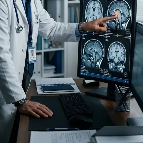
Do not wait to see whether the symptoms settle. A blocked artery causes an ischaemic stroke; bleeding causes a haemorrhagic stroke, and aspirin can worsen a brain haemorrhage. At hospital, doctors usually begin with a non-contrast CT head to detect bleeding, followed by CT angiography to examine blood vessels.
CT perfusion is added when indicated, while MRI helps in selected cases. A mild early examination does not rule out a dangerous blocked large vessel.
How Hospitals Identify the Stroke and Choose Emergency Treatment
A non-contrast CT head is the first critical scan: it detects bleeding and shows whether a large area is already irreversibly injured. An ischaemic stroke caused by a blocked artery needs a different plan from a haemorrhagic stroke caused by bleeding.
Record the exact symptom-onset time, or the last known well if nobody saw the onset, and alert 112 emergency services.
A contrast CT head, usually CT angiography, looks for a blocked major artery. CT perfusion measures salvageable brain tissue; MRI helps in selected cases when the onset time is unclear or treatment is considered late.
| Treatment | What doctors assess | Possible action |
|---|---|---|
| Intravenous thrombolysis | Disabling ischaemic stroke, haemorrhage excluded, treatment within 4.5 hours | Clot-dissolving medicine after checking contraindications, blood pressure, and timing |
| Mechanical thrombectomy | Major artery occlusion, limited established infarction, suitable function and imaging | Catheter clot removal, up to 24 hours in selected patients |
| Intensive medical treatment | No eligible clot procedure, smaller occlusion, or bleeding stroke | Blood-pressure control, monitoring, stroke-unit care, and complication prevention |
| Decompressive hemicraniectomy | Life-threatening swelling after a large infarction | Skull-pressure relief in carefully selected patients; survival does not guarantee restored function |
Do not give aspirin before imaging: aspirin is a danger in haemorrhage. A normal early examination does not exclude a major-vessel blockage, and MRI or CT perfusion can change the decision when symptoms, timing, and scans disagree.
When Stroke Surgery Is Needed—and What It Can and Cannot Repair
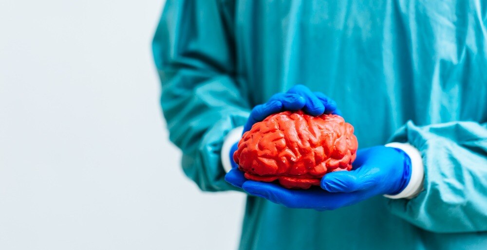
An operation is needed when swelling or bleeding threatens the brain, not simply because a stroke caused weakness. A large middle-cerebral-artery infarction with dangerous brain swelling may require decompressive craniectomy, usually a decompressive hemicraniectomy, to reduce fatal pressure.
A swollen cerebellar infarction can cause brainstem compression or obstructive hydrocephalus; suboccipital decompression, sometimes with external ventricular drainage, should not wait for profound deterioration.
An intracerebral haematoma may need intracerebral haematoma evacuation when its size, location, or pressure causes deterioration. In aneurysmal subarachnoid haemorrhage, the ruptured aneurysm is secured with clipping or endovascular coiling to prevent rebleeding.
| Option | What it does | When it applies |
|---|---|---|
| Medical care | Controls pressure, oxygenation, glucose, seizures, and other complications | Most strokes and patients unsuitable for procedures |
| Catheter treatment | Mechanical thrombectomy removes an accessible artery clot without opening the skull | Selected large-vessel ischaemic stroke, sometimes up to 24 hours with salvageable tissue |
| Surgery | Relieves pressure, removes selected blood, or secures an aneurysm | Dangerous swelling, haemorrhage, or aneurysmal subarachnoid haemorrhage |
Surgery can reduce death but cannot reverse already injured brain tissue. Paralysis, speech loss, memory and visual deficits may remain, making rehabilitation essential. Minimally invasive or endoscopic brain surgery has specific access advantages, but its limitations mean it cannot replace decompression or thrombectomy when those are indicated.
Dr. Deepak Bariya can help families understand which option matches the scan, timing, and neurological examination.
Related service
What Is a Stroke (CVA)? A stroke happens when part of the brain does not receive enough blood or oxygen. View service → |
How to Compare Jaipur Stroke Centres and Prepare for Specialist Assessment
Check whether a Jaipur hospital provides 24-hour CT, MRI, and CT angiography, plus neuro-intensive care, neurosurgery, interventional neuroradiology, thrombectomy, anaesthesia, blood-bank support, and rehabilitation. Confirm its emergency transfer pathway; a listing that says “neurosurgery” does not prove round-the-clock stroke capability.
| Centre feature | Why it matters | Question to ask |
|---|---|---|
| Local stroke unit | Stabilises patients and completes urgent imaging | Is a stroke team present now? |
| Thrombectomy service | Enables catheter treatment for selected blockages | Is interventional neuroradiology available 24 hours? |
| Surgical backup | Treats swelling, bleeding, or hydrocephalus | Can neurosurgery and anaesthesia respond immediately? |
Bring these details for specialist assessment:
- The exact neurological examination findings, blood pressure, age, frailty, diabetes, kidney disease, allergies, recent surgery, and prior scans.
- The anticoagulant name and last dose, platelet count, and clotting tests.
- The symptom-onset or last-known-well time, current medicines, and previous medical records.
Ask whether private specialist consultation after stabilisation with Dr. Deepak Bariya can be arranged. Before discharge, obtain imaging reports, CT/MRI copies, the confirmed stroke mechanism, medication plan, warning signs, and rehabilitation follow-up instructions.
What Happens After Emergency Treatment and During Rehabilitation
After emergency treatment, expect neuro-intensive monitoring of consciousness, pupils, strength, oxygen levels, heart rhythm, temperature, and blood pressure. Repeat CT scans are used when doctors need to check bleeding, swelling, hydrocephalus, or a change in neurological function; a sudden decline requires immediate reassessment.
Food, drink, and oral medicines must wait until a swallowing assessment is passed. Stroke-related dysphagia can cause silent aspiration without coughing, so aspiration prevention may include supervised sips, upright positioning, altered textures, oral care, or temporary tube feeding. The team will explain how oral intake and nutrition will be provided safely.
Once medically stable, early rehabilitation begins, but very high-intensity mobilisation during the first 24 hours is not routinely suitable and can harm some patients. Families should expect a coordinated plan involving:
- Physiotherapy to improve movement, balance, transfers, and walking.
- Occupational therapy for dressing, washing, hand use, and home activities.
- Speech and language therapy for speech, communication, and swallowing.
- Cognitive and psychological support for attention, memory, mood, behaviour, and adjustment.
Nursing care also covers blood-clot prevention, pressure-injury prevention, bladder and bowel needs, and blood-pressure management. Ask which exercises are safe, what supervision is required, and how progress will be measured before attempting practice at home.
Planning Discharge, Recovery, and Prevention of Another Stroke
Before discharge, obtain the imaging reports, CT and MRI images, confirmed stroke mechanism, medication plan, warning signs, wound or drain instructions, and a written follow-up and rehabilitation schedule. Confirm who will provide caregiver supervision, physiotherapy, speech therapy, and help with loss of independence; discuss practical care costs before transport home.
- Start blood-pressure monitoring, diabetes control, cholesterol control, smoking cessation, and atrial fibrillation evaluation. Record whether antiplatelet or anticoagulant decisions are complete; never change either drug without the stroke team.
| Change | Action | Why |
|---|---|---|
| Worsening headache, repeated vomiting, new weakness, confusion, seizure, or reduced alertness | Call emergency services immediately | Could signal bleeding, swelling, or another stroke |
| Speech deterioration or new facial droop | Note the last-known-well time and seek emergency transport | Improvement later does not make it safe to observe |
| Fever, wound leakage, or a displaced drain | Contact the surgical team urgently | Infection or pressure complications need assessment |
Frequently asked questions
What are the first signs of a stroke?
Use FAST: face drooping, arm weakness, speech difficulty, and time to call emergency services. Sudden confusion, leg weakness, vision loss, severe imbalance, seizure, or an abrupt severe headache also require immediate help.
How do hospitals diagnose a stroke and choose treatment?
The emergency team assesses neurological symptoms and uses brain imaging to determine whether the stroke involves bleeding, a blocked blood vessel, swelling, or another cause. Those findings guide emergency treatment.
When is stroke surgery needed?
Surgery is considered when bleeding, pressure, swelling, or a blocked blood vessel creates a serious risk that requires an intervention. The treating specialist explains what the procedure can address and what brain damage it cannot reverse.
What should you check when comparing stroke centres in Jaipur?
Ask whether the centre provides emergency brain imaging, specialist neurosurgical assessment, intensive care, stroke treatment, rehabilitation planning, and follow-up support.