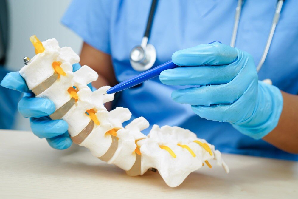
Leg pain, numbness, or weakness does not automatically mean that an MRI finding needs surgery. You will learn how surgeons match symptoms to imaging, select the safest treatment and endoscopic route, and compare the practical capabilities of spine hospitals in Jaipur.
Key takeaways
- Match leg symptoms, examination findings, and MRI evidence before considering surgery.
- Ask why an endoscopic route fits your anatomy and which alternatives remain available.
- Complete medication review, medical tests, and surgical planning before the operation.
- Compare surgeon experience, hospital support, follow-up access, and complication planning.
Who is a suitable candidate for endoscopic spine surgery?
Suitable endoscopic spine surgery candidates have leg-dominant pain, numbness, or weakness caused by a single compressed lumbar nerve, with symptoms that match the neurological examination and MRI. The clearest example is lumbar disc herniation producing sciatica after medication, activity modification, physiotherapy, or observation has not provided enough improvement.
An abnormal MRI without matching symptoms is not a reason to operate.
| MRI and clinical findings | When endoscopic decompression is reasonable | When another priority takes over |
|---|---|---|
| Focal lumbar disc herniation | Leg pain follows a nerve-root pattern, and MRI shows a fragment compressing that root | Back pain alone, no neurological match, or symptoms improving with non-operative care |
| Foraminal stenosis | A narrowed neural foramen visibly compresses the exiting nerve and symptoms fit | Severe bony narrowing requiring wider decompression or fusion |
| Lateral recess stenosis | The traversing nerve is compressed in the lateral recess, with persistent radicular pain or weakness | Extensive central stenosis, deformity, or instability |
| Recurrent disc herniation or selected spinal stenosis | Repeat imaging identifies a focal, reachable compression | Infection, tumour, fracture, significant instability, or a case requiring fusion |
Do not wait for a routine treatment course if weakness is progressing, neurological loss is severe, or cauda-equina symptoms appear. New urinary retention or incontinence, loss of bowel control, saddle numbness, or rapidly worsening weakness needs urgent assessment.
A large incision is not automatically better, but endoscopy is not suitable for every compression; microscopic or open decompression, injection treatment, physiotherapy, or continued observation may provide the safer choice.
How do surgeons choose the route and the alternative treatment?
The choice starts with MRI anatomy and neurological findings, not incision size. The surgeon checks the disc fragment’s level, location and migration, foraminal height, facet anatomy, instability, deformity and prior operations, then matches the route to the compressed nerve.
| Option | What it means | When it applies |
|---|---|---|
| Transforaminal endoscopic spine surgery | Enters through the neural foramen | Foraminal or lateral disc compression and selected migrated fragments |
| Interlaminar endoscopic spine surgery | Enters between the laminae | Central or lateral recess compression when the interlaminar window is suitable |
| Microscopic decompression | Removes bone or disc through a microscope | Compression needing wider access than an endoscope provides |
| Open surgery or fusion | Decompresses and stabilises the spine | Instability, major deformity, fracture, tumour, infection, severe central stenosis or multilevel disease |
| Injection treatment | Places medication near an irritated nerve | Temporary relief or diagnostic clarification without an urgent neurological deficit |
| Physiotherapy | Builds strength and changes spinal loading | Mechanical pain, stable symptoms and recovery without progressive weakness |
| Observation | Monitors symptoms and examination | Improving symptoms without significant neurological loss |
Endoscopic access is not automatically safer or more effective. A transforaminal route can irritate the exiting nerve root, while an interlaminar route works near the traversing root and dura. Severe central stenosis or a case requiring fusion can make conventional surgery the safer spinal stenosis treatment.
For uncomplicated sciatica, injections, physiotherapy or observation usually come before surgery. Progressive weakness, worsening neurological loss or cauda-equina symptoms require urgent assessment. Isolated back pain without convincing nerve compression also makes decompression less predictable.
What happens before the operation?
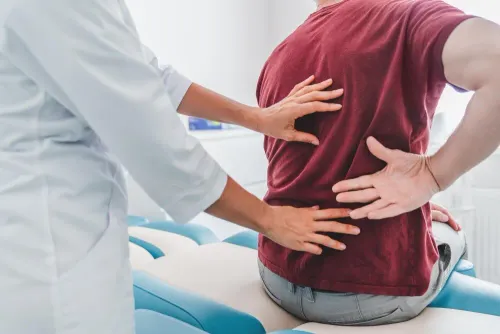
The decision starts with a history of pain location, duration, walking limits, numbness, weakness, bladder or bowel change, previous treatment, earlier spine surgery, medicines, allergies, diabetes, heart or lung disease, and infection risk. Progressive weakness or cauda-equina symptoms require urgent assessment rather than a routine surgical pathway.
The clinical review should include:
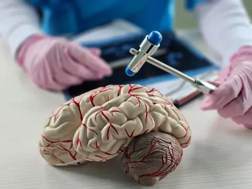
- Strength, sensation, reflexes, gait, straight-leg raising, and signs of nerve-root tension.
- MRI review for spine surgery, checking the symptomatic level, disc fragment, migration, foraminal height, lateral recess, central canal, facet joints, and prior surgical changes.
- A standing X-ray spine to assess alignment, vertebral height, disc-space narrowing, deformity, and suspected instability.
- Flexion-extension X-rays when movement-related instability or spondylolisthesis could change the operation.
- Blood tests such as a complete blood count, kidney function, blood glucose, electrolytes, and coagulation tests when indicated by medical history or planned anaesthesia.
| Test or discussion | What it establishes | Why it matters |
|---|---|---|
| MRI | A compressing disc or narrowed nerve passage matching the examination | An abnormal scan without matching symptoms is not enough |
| Standing and movement X-rays | Alignment and instability under load | Instability can favour decompression with fusion or another approach |
| Preoperative assessment | Fitness for anaesthesia, medication risks, airway, and recovery needs | Uncontrolled medical problems can delay surgery |
| Anaesthetic discussion | Local, regional, or general anaesthesia plan; fasting and medicine instructions | It clarifies pain control, monitoring, and safety during the procedure |
What happens during recovery, and what risks should you understand?
The operation usually follows a short, controlled sequence:
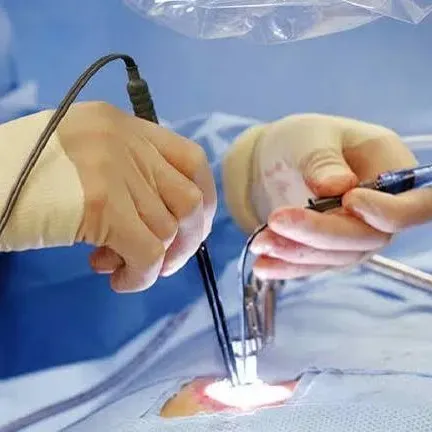
1. You receive positioning, skin preparation, and fluoroscopic imaging to confirm the spinal level. The surgeon makes a small incision, passes a working tube, and uses irrigation and an endoscope to reach the nerve or disc.
2. The surgeon removes the disc fragment or bone causing compression, checks nerve-root movement, withdraws the instruments, and closes the incision. Staff then monitor leg strength, sensation, pain, blood pressure, and urination before discharge or admission.
3. Local anaesthesia with sedation can keep you responsive during some lumbar procedures, so you may feel pressure without sharp pain. General anaesthesia spine surgery means you are fully unconscious; it may suit a long, painful, technically difficult, revision, cervical, thoracic, or multilevel operation.
Endoscopic discectomy recovery often involves walking on the day of surgery, wound care, and avoiding bending, twisting, heavy lifting, and driving until your surgeon permits them. Increase activity gradually; do not judge the result by pain alone during the first days.
Endoscopic spine surgery risks include infection, bleeding, nerve injury, dural tear with cerebrospinal-fluid leakage, incomplete decompression, anaesthetic complications, and conversion to conventional surgery. A removed disc fragment does not reverse disc degeneration, so recurrent herniation or persistent symptoms require examination and, when indicated, repeat imaging.
Seek emergency care for new urinary retention, loss of bladder sensation, faecal incontinence, saddle-area numbness, or rapidly worsening weakness.
How can you compare endoscopic spine care in Jaipur?
Do not compare clinics by the word “minimally invasive”; compare the team’s ability to select, perform, and safely support the operation. An endoscopic spine surgeon Jaipur patients consider should explain why the transforaminal or interlaminar route fits your MRI, what alternative he or she recommends, and when endoscopy is the wrong choice.
Dr. Deepak Bariya is one surgeon you can assess using these same practical questions.
| Criterion | What to verify | Warning sign |
|---|---|---|
| Surgeon | Training, experience with your diagnosis, and a clear plan if decompression is inadequate | Promises of guaranteed results |
| Hospital | A spine surgery hospital Jaipur with trained anaesthesia, operating-room staff, sterilisation, and irrigation systems | Procedure offered without clear inpatient support |
| Imaging | Real-time fluoroscopy spine surgery capability and equipment suited to the planned route | Vague answers about image guidance |
| Emergency readiness | Management of bleeding, dural injury, nerve injury, infection, and conversion to conventional surgery | No named escalation plan or emergency access |
| Recovery | Postoperative neurological observation, discharge criteria, and access to urgent review | Same-day discharge with no contact pathway |
Before choosing, ask:
- Who will perform the operation, and how often do they use the proposed route?
- What finding on my MRI matches my symptoms?
- What happens if the fragment cannot be reached or the nerve remains compressed?
- Who answers after discharge if weakness, fever, worsening pain, or bladder symptoms appear?
- When are wound review, neurological review, and repeat imaging required?
Related services
Endoscopic Spine Surgery( Slip Disc Surgery) What Is Endoscopic Spine Surgery? Endoscopic spine surgery is a minimally invasive procedure performed using a small endoscope—a thin tube with a... View service → |
Headache And Back Ache Minimally Invasive Brain & Spine Surgery ₹500 What Is Minimally Invasive Brain & Spine Surgery? Minimally Invasive Brain & Spine Surgery (MIS) involves performing surgical procedures through very... View service → |
Frequently asked questions
Who is a suitable candidate for endoscopic spine surgery?
The clearest candidates have leg-dominant pain, numbness, or weakness from a single compressed lumbar nerve, with symptoms matching the neurological examination and MRI. Surgery is considered after medication, activity modification, physiotherapy, or observation has not provided enough improvement.
How do surgeons choose the route and alternative treatment?
The surgeon matches the endoscopic route to the disc location, level, spinal anatomy, nerve compression, and diagnosis. They also compare alternatives such as continued non-surgical care, injections, conventional decompression, or fusion when instability or another structural problem makes endoscopy unsuitable.
What happens before endoscopic spine surgery?
Before surgery, the team reviews your symptoms, neurological examination, MRI findings, medicines, allergies, medical conditions, and previous treatments. You may need blood tests, anaesthesia assessment, medication instructions, and specific guidance about fasting, transport, and post-operative support.
What happens during recovery, and what risks should you understand?
Recovery usually focuses on wound care, walking, pain control, activity limits, and follow-up assessment of leg symptoms. Ask about recurrent disc herniation, infection, bleeding, nerve injury, dural tear, persistent symptoms, and the signs that require urgent medical attention.
How can you compare endoscopic spine care in Jaipur?
Compare the surgeon’s training and experience with your diagnosis, the hospital’s anaesthesia and emergency support, the proposed technique, alternatives offered, expected follow-up, total treatment plan, and how complications would be managed.