A brain lesion on an MRI does not by itself establish a tumour type or determine whether surgery is needed. You need to understand how doctors confirm the diagnosis, how location changes the surgical plan, what “maximal safe resection” means, and which questions to ask before consenting.
Key takeaways
- MRI shows the lesion; tissue analysis confirms its tumour type.
- Ask why observation, biopsy, surgery, or another treatment fits your case.
- Tumour location determines the surgical route and risks to speech, movement, or vision.
- Bring scans, medication details, symptoms, and questions to your consultation.
How doctors confirm a brain tumour diagnosis
A neurological examination checks strength, sensation, coordination, vision, speech, memory, and balance. These findings help doctors match symptoms to the abnormality’s location, but they cannot prove that the lesion is a tumour.
A contrast-enhanced MRI usually provides the clearest view of the lesion, its borders, swelling, blood supply, and relationship to normal brain. A CT scan for brain tumour surgery adds useful information about bleeding, calcification, bone involvement, hydrocephalus, or an emergency when MRI is unavailable.
Doctors may add these tests when MRI shows a lesion near important brain networks:
- Functional MRI maps language or movement areas before an operation.
- Tractography follows nerve-fibre pathways that carry movement, vision, or language signals.
- Neuropsychological testing measures memory, attention, language, and other functions as a baseline.
- A follow-up scan may show whether an abnormality changes like a tumour or behaves more like inflammation.
Imaging can suggest a meningioma, glioma, metastasis, abscess, haemorrhage, or demyelinating disease, but appearances overlap. A brain tumour is not automatically cancer, and even a benign tumour can obstruct cerebrospinal-fluid flow or compress vital tissue.
A biopsy or tissue obtained during surgery is often needed for a definitive brain tumour diagnosis. A neuropathologist examines the cells and uses molecular tests to identify the tumour type, grade, and features that guide treatment. Tissue sampling is chosen only after weighing the lesion’s location and the risks of reaching it.
When observation, biopsy, or treatment is the right next step
Observation is reasonable for a small, symptom-free lesion that has benign imaging features and remains stable on scheduled MRI scans. Doctors recommend tissue diagnosis or treatment when the lesion grows, causes seizures or pressure, produces neurological symptoms, or has imaging features that could represent a high-grade tumour, abscess, metastasis, or another disease.
| Option | What it means | When it applies |
|---|---|---|
| Observation | Repeat MRI and clinical review | Stable, low-risk lesions without significant symptoms |
| Brain tumour biopsy | Removes tissue for microscopic and molecular testing | The diagnosis will change treatment and removal is unsafe or unnecessary |
| Stereotactic needle biopsy | Uses image guidance to sample a deep or difficult lesion through a small opening | A large craniotomy carries greater risk; a small sample can miss aggressive tumour or provide insufficient molecular material |
| Gross-total resection | Removes all visible tumour | The tumour is accessible and removal can preserve speech, movement, vision, memory, and other functions |
| Subtotal resection | Leaves tumour deliberately to protect critical brain tissue | The lesion involves an eloquent area, deep structure, or vital blood vessels |
| Radiotherapy or chemotherapy | Treats remaining or infiltrative tumour cells | Pathology, grade, recurrence risk, or tumour spread supports additional treatment |
| Targeted or combined treatment | Uses a molecularly matched drug with surgery, radiation, or chemotherapy | Molecular results identify a treatable alteration or high-grade disease needs several treatments |
A multidisciplinary team uses MRI findings, pathology, molecular results, symptoms, age, general health, and functional mapping to set the plan. Corticosteroids can reduce swelling, while anti-seizure medicine treats seizures; neither replaces tumour-directed treatment.
Why tumour location changes the operation
A tumour beside speech or movement networks can make complete removal more dangerous than leaving a thin margin. The goal becomes maximal safe resection: removing as much tumour as possible while preserving the functions that control independence and communication.
Planning uses contrast-enhanced MRI and neuronavigation to locate the lesion during surgery. Functional MRI, tractography, or neuropsychological testing can show nearby networks, but none guarantees normal function after removal.
- Near speech areas, surgeons may use an awake craniotomy. The patient is awake during testing while the team uses electrical stimulation for cortical mapping, identifying tissue that supports language before removing tumour. This approach is unsuitable for some patients.
- Near movement pathways, stimulation and careful monitoring help identify fibres controlling the face, arm, or leg. A planned subtotal resection can be safer than causing permanent weakness.
- Near visual or memory networks, the surgeon weighs the risk of visual-field loss or cognitive change against the benefit of removing more tissue.
- Near swallowing pathways or the brainstem, even a small injury can affect swallowing, breathing, balance, or consciousness, so biopsy or limited removal may replace aggressive resection.
- In the skull base or deep brain, narrow access routes and major blood vessels restrict the angle of surgery. A ventricular tumour may obstruct cerebrospinal-fluid flow, making drainage or removal necessary to relieve pressure while protecting nearby structures.
Tumour position also affects risks such as bleeding, seizures, swelling, fluid leakage, and new neurological deficits. Ask which function is at risk, how it will be tested, and what amount of tumour removal is realistically safe.
What happens during surgery and recovery
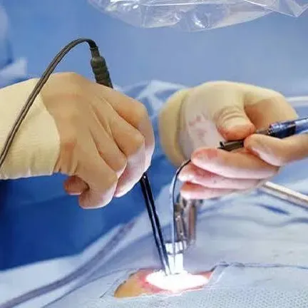
Surgeons choose the route that reaches the tumour while protecting speech, movement, vision and memory networks. Endoscopic brain surgery uses a narrow camera through a small opening for selected tumours, especially in or near fluid-filled spaces. Intraoperative MRI can update tumour images during the operation, while neurophysiological monitoring tracks vulnerable nerve pathways.
Craniotomy versus craniectomy matters: a craniotomy replaces the bone flap before closure; a craniectomy leaves it out temporarily when swelling makes replacement unsafe, requiring later reconstruction. The operation can cause:
- Bleeding, infection, seizures or cerebrospinal-fluid leakage
- Brain swelling, blood clots or anaesthetic complications
- New or worsened weakness, speech difficulty, visual loss or cognitive change
Ask for outcome data from operations involving your tumour’s location, not one overall complication rate.
A postoperative MRI is commonly performed soon after resection to measure residual enhancing tumour and establish a baseline. Blood products and inflammation can resemble tumour on an early scan, so timing, technique and later comparison affect interpretation.
Your discharge plan may include dexamethasone for swelling, an antiseizure medicine, pain relief and clot-prevention measures. Take medicines exactly as prescribed and attend physiotherapy, occupational therapy or speech therapy when recommended; temporary deficits can improve, but some remain permanent.
Contact the surgical team urgently or seek emergency care for:
- Repeated vomiting, worsening headache, drowsiness or confusion
- A new seizure, weakness or speech trouble
- Fever, wound redness, drainage or clear fluid from the wound or nose
How to prepare for consultation and consent in Jaipur
Bring MRI images and previous scans on a viewer or digital link, radiology reports, medication and seizure records, blood-test results, and any biopsy slides or reports.
Ask a brain tumour surgeon in Jaipur to state the working diagnosis, alternatives, intended surgical endpoint, functions at risk, likelihood of biopsy or residual tumour, and expected hospital stay and activity restrictions.
Before signing consent, ask:
- How will MRI with contrast, functional MRI, tractography, or neuropsychological testing change the plan?
- Who will review pathology and molecular testing, and when will those results alter treatment?
- Who coordinates radiotherapy, oncology, seizure treatment, steroids, rehabilitation, and follow-up?
- What are this centre’s outcomes for operations in the relevant brain region, including temporary and permanent neurological deficits?
| Plan | What to compare | Main trade-off |
|---|---|---|
| Maximal safe resection | Expected tumour removal and function risk | More tissue diagnosis and control versus greater risk near speech, movement, vision, or memory pathways |
| Needle or open biopsy | Diagnostic yield and access route | Less removal than resection, but treatment still follows |
| Observation or non-surgical treatment | Scan schedule and trigger for intervention | Avoids an operation but requires reliable monitoring |
A second neurosurgical opinion is reasonable for elective surgery, uncertain diagnosis, or disagreement about the endpoint. Dr. Deepak Bariya can help you frame that discussion around imaging, tumour location, centre-specific outcomes, and the complete treatment team rather than surgical technique alone.
Frequently asked questions
How do doctors confirm a brain tumour diagnosis?
Doctors combine neurological examination with brain imaging such as MRI or CT. Imaging shows the lesion’s size and location, while biopsy or tissue removed during surgery can identify its tumour type and grade.
When is observation, biopsy, or treatment the right next step?
The choice depends on the lesion’s appearance, growth, symptoms, location, and suspected tumour type. Small, slow-growing lesions without symptoms may be monitored; tissue sampling or treatment is considered when diagnosis or symptom control requires it.
Why does tumour location change the operation?
A tumour near speech, movement, vision, memory, or vital blood vessels requires a surgical plan that protects those functions. Location also affects the safest route and how much tumour can be removed.
What happens during brain tumour surgery and recovery?
The surgical team uses imaging and neurological monitoring to remove or sample the lesion. Recovery depends on the operation and your neurological status, with hospital observation, symptom management, pathology review, and follow-up planning.